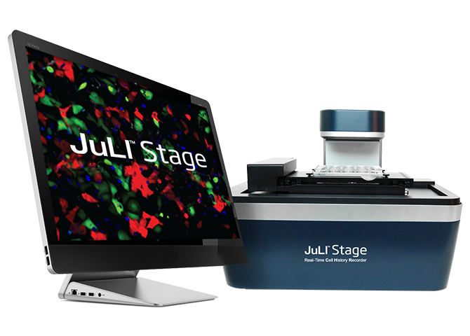
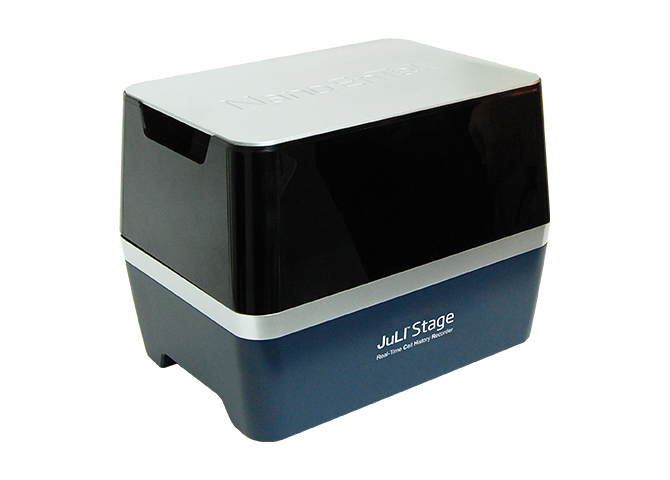
JuLI™ Stage Live Cell Imager is a compact live cell imaging system ready fit into almost any CO2-incubator. JuLI™ Stage offers automated X-, Y- and Z-axis control, interchangeable objectives with 4x, 10x and 20x zoom and Multiple light sources (Bright Field, Green-, Red- and Blue fluorescent channels).
The automated axis control and fluorescent channels along with the Cell History Recorder (CHR) software allows the JuLI™ Stage to automate procedures such as image capturing, Time-Lapse, video making, image stitching, multi-well monitoring and Multi position monitoring.
All ideal for investigating:
• Wound healing
• Apoptosis & Cytotoxicity
• Cell proliferation
• Fluorescence expression
• 3D-spheroid culture
• Stem cell monitoring
• Cell growth monitoring
• Differentiation of neurons
• Angiogenesis
To Fully automate Scratch assays we also offer JuLI Scratch STAT and JuLI Scratcher.
If you work with Spheriods we can supply a special Spheroid STAT software.
Scope of supply: Real-time live cell imaging system, desktop computer, desktop monitor, 3 objective lenses (4x, 10x and 20x), CHR software, EDIT software, STAT software.