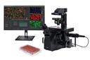
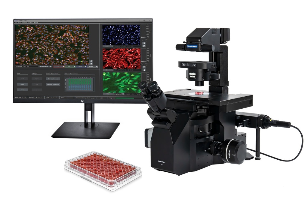
High-Content Screening Solution: Olympus IX85 scanR
Much more than just high-content screening: flexible, modular, and robust hardware
The scanR screening station combines the modularity and flexibility of a microscope-based setup with the automation, speed, throughput, and reproducibility needed for high-content screening applications. The system is designed for a range of applications, including standard assays and assay development, and its modularity makes the scanR station adaptable for R&D lab applications and multiuser environments. The scanR system features sophisticated image-analysis and data-analysis software that uses an interactive, cytometry-oriented workflow, enabling it to analyze large numbers of multidimensional data sets.
Designed for fully automated image acquisition and data analysis, the scanR solution accomodates multiwell plates, slides, and custom-built arrays. The system can handle fixed and live cells, and the screening station specifically targets the requirements for quantitative imag analysis in modern cell biology, molecular biology, systems biology, and medical research.
A scanR screening station can be built on multiple platforms. The most versatile solution is the new IX85 inverted microscope for a fully motorized, modular setup allowing for future expansion. We can also build your scanR as:
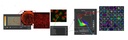
- Spinning Disc Confocal - Acquire high-resolution, high-contrast images using the IXplore SpinSR super resolution microscope that includes the Yokogawa CSU-W1 scanner unit. Micro-lens-based disks and laser excitation provide seamless confocal image quality at high speed.
- Robot Loading System Setup - For automated high-throughput screening, the scanR system can be combined with a plate-loading robot.
- Incubation System Setup - Combining the scanR high-content screening solution with an incubation system provides strict control of temperature, humidity and CO2.
- TIRF and FRAP System Setups - The scanR platform is compatible with the IXplore Family of microscopes, which, combined with cellSens software, enables users to perform advanced imaging experiments such as TIRF and FRAP.
Highlights:
Set up your analysis during acquisition - Most of the analysis features are available on the fly, enabling users to perform immediate quality control and generate statistics in just a few seconds.
Clear guidance - Easy workflow provides reliable image acquisition and straightforward system configuration for accurate, repeatable quantitative measurements.
Maintain your focus - Fast and accurate autofocus maintains the focus plane using a combination of software algorithms and hardware.
More dimensions - The system’s advanced features enable truly multidimensional (X, Y, Z, λ) screening. Record time-lapse Z-stacks at numerous locations, using all available observation methods (fluorescence, brightfield, differential interference contrast (DIC), and phase contrast).
Optimized for X Line objectives - Outstanding image quality is a fundamental requirement for quantification. The scanR system supports Evident X Line objectives to deliver broad chromatic aberration correction, uniform images, and a high numerical aperture (NA).
Works with almost any well or slide - The system is adaptable to any standard well plate format or slide, and is capable of being calibrated to any regular pattern, such as spotted arrays.
Multilevel acquisition - Based on an initial prescan, the scanR analysis software can identify all the potential objects of interest. In an automated workflow, the analysis results are used to selectively scan the objects of interest in a second, targeted screen. This multilevel acquisition is especially beneficial for single-cell events or high-resolution imaging of large-area samples with few cells.
Examples of cellular screening assays:
- Cell viability
- Gene expression
- Intracellular transport
- Translocation
- Cell proliferation
- Promyelocytic leukemia (PML) body assay
- Bacterial and viral infection assays
- Cell-cycle analysis
- Cell-array screens
- Multicolor assays
- Rare-event analysis
- Automated-FISH analysis
- Fluorescence analysis in tissue sections
- Live-cell assays including kinetic analysis and gating on resulting response curves
- Micronuclei and comet assays
- Cell migration
- Protein localization and colocalization